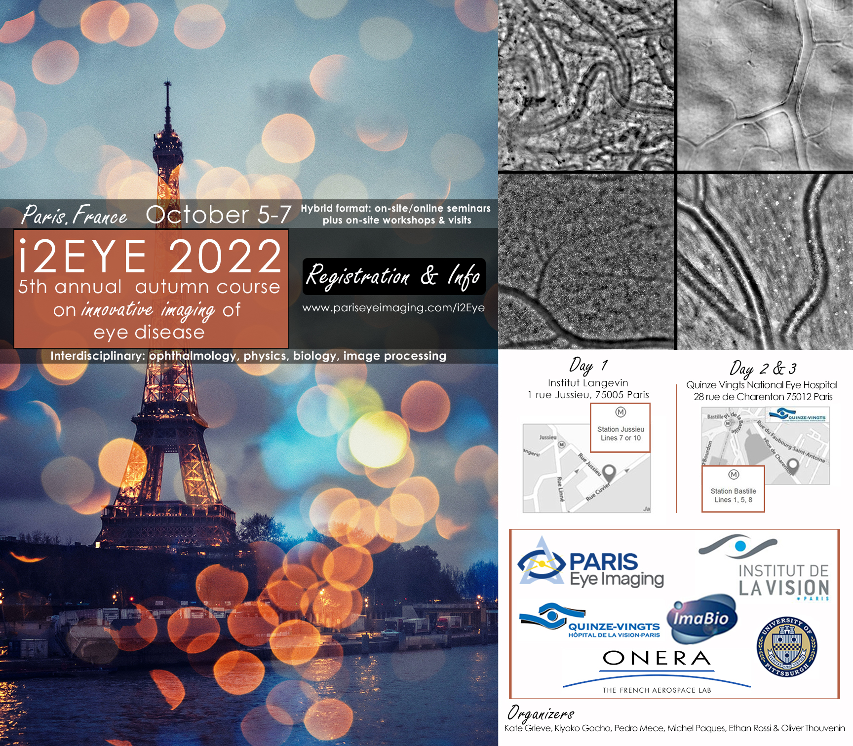
Innovative Imaging of Eye disease (5 - 7 October 2022)
All of the presentations can be downloaded here:
Day 1 - October 5
Session 0: Introductory courses - Moderator: K Grieve Download video of Session 0
The eye - clinical viewpoint - Michel Paques
Imaging the eye in vivo - Pedro Mecê
The eye – biology viewpoint - Anna Verschueren
Microscopy - Olivier Thouvenin
Session 1: Novel technology - Moderator: P MecêDownload video of Session 1
Keynote: Multimodal retinal tissue assessment combining wide field OCT/OCTA and Raman Spectroscopy - Rainer Leitgeb
Revealing cells in the retina with “transmission” microscopy in vivo - Elena Gofas, Nate Norberg, Michel Paques, Kate Grieve
Multimodal and multiscale clinical high-resolution retinal imaging - Kiyoko Gocho, Céline Chaumette, Kate Grieve, Nicolas Lefaudeux, Marin Cor, Marine Durand, Xavier Levecq, Nicolas Chateau, Michael Pircher, Michel Paques
Multi-aperture AO-SLO retinal imaging - Mircea Mujat, Ankit Patel, Nicusor Iftimia
High resolution pattern projection in the retina for phase contrast imaging - Pierre Senée, Léa Krafft, Pedro Mecê, Serge Meimon
Laser holography for ophthalmology - Michael Atlan
Multi-spectral matrix microscope - Paul Balondrade, Victor Barolle, Nicolas Guigui, Claude Boccara, Mathias Fink, Alexandre Aubry
Discussion
Session 2: Histology, organoids and disease models - Moderator: E Gofas Download video of Session 2
Keynote: Creating retinal organoids: new insights on human development and diseases - Olivier Coureau
Timelapse imaing of retinal organoids and cell cultures with dynamic FFOCT microscopy - Tual Monfort, Salvatore Azzollini, Olivier Thouvenin, Kate Grieve
Comparing clinical features with hitology - Michel Paques, Anna Verschueren, Marie Darche
Dynamic OCT with retinal pigment epithelium-choroid tissue ex-vivo - Yoko Miura, Tabea Kohlfaerver, Niroko Kubota, Martin Ahrens, Mario Pieper, Peter König, Gereon Hüttmann, Hinnerk Schulz-Hildebrandt
Three-dimensional imaging and quantitation of the collagen lamellae within the human cornea using polarimetric SHG imaging - Clothide Raoux, Anatole Chessel, Marie-Claire-Schanne-Klein, Caël Latour
Discussion
Day 2 - October 6
Session 3: Functional Imaging and Eye Movements - Moderator: J Morgan Download video of Session 3
Keynote: optoretinography and retinal imaging in retinal health and disease using adaptive optics scanning laser ophthalmoscopy - Jessica Morgan
All-optical inter-layers functional connectivity investigation in the mouse retina - Emiliano Ronzitti, GLB Spampinato, V Zampini, UFerra, F Trapani, H Khabou, A Agraval, D Dalkara, S Picaud, E Papagiakoumou, O Marre, V Emiliani
Does manipulation of retinal slip during fixational drift impacts visual acuity? - Veronika Lukyanova
Characterization of retinal axial motion and its implications for real-time correction in in vivo human retinal imaging - Yao Cai, Olivier Thouvenin, Kate Grieve, Pedro Mecê
Optoretinography of the inner plexiform layer - Clara Pfäffle, Svea HöHl, Hendrik Spahr, Léo Puyo, Jonas Francke, Dierck Hillmann, Cereon Hüttmann
Optoretinography measurement of human retina response to flickering stimuli - Slawomir Tomczewski, Piotr Wegrzyn, Dawind Borycki, Maciej Wielgo, Andrea Curatolo, Maciej Wojtkowski
Fixational eye movements measured via AOSLO – do system and processing effects matter? - Laura Young
Discussion
Session 4: Anterior segment and glaucoma - Moderator: K Gocho Download video of Session 4
Detection of stages of glaucoma through image processing - Syeda Fizza Batool
Assessment of corneal biomechanical asymmetry via differential multi-spot air-puff OCT measurements - Karol Karnowski, Jadwiga Milkiewicz, Angela Pachacz, Andrea Curatolo, Onur Cetinkaya, Rafal Pietruch, Alejandra Consejo, Maciej M Bartuzel, Piotr Ciacka, Ashkan Abass, Ahmed Elsheikh, Susana Marcos, Maciej Wojtkowski
Compact cell-resolution anterior eye imager: technology and the first clinical cases - Slava Mazlin
Discussion
Clinical case share: impromptu 5 minutes slots to show and discuss your clinical cases and questions concerning high resolution imaging technology
Day 3 - October 7
Session 5: Retinal cellular biomarkers - Moderator: R Baraas Download video of Session 5
Keynote: Between-individual variation in human macular cone and RPE cell topography - Rigmor Baraas
Physiopathology of dry AMD: deciphering the cellular dynamics underlying progression of atrophy - Ysé Borella, A Verschueren, M Darche, K Grieve, F Sennlaub, N Norberg, J Gautier, E Rossi, J-A Sahel, F Rossant, C Chaumette, M Paques
Loss of cone outer-segments in multiple sclerosis: The Belfast eye and Multiple Sclerosis Study - Gemma McIlwaine, Lajos Csincsik, Rachel Coey, Luping Wang, Denise Fitzgerald, Jill Moffatt, Gavin McDonnell, Stella Hughes, Adam Dubis, Tunde Peto, Imre Lengyel
Adaptive optics retinal imaging in choroideremia - Katarina Stingl
Label-free in vivo imaging of inflammation at the level of single cells in the living human eye - Ethan Rossi
Adaptive Optics in Non-Human Primates: normal and induced pathological findings - Alexandre Dentel, C Jaillard, K Grieve, M Paques, S Bertin, E Brazhnikova, V Fradot, S Picaud
Discussion
Session 6: Blood Flow and Vasculature - Moderator: M Paques Download video of Session 6
In vivo imaging of choroid by Spatio-Temporal Optical Coherence Tomography - Kamil Lizewski, Egidijus Auksorius, Piotr Wegrzyn, Mounika Rapolu, Leva Zickiene, Karolis Adomavicius, Slawomir Tomczewski, Bartosz L Sikorski, Dawid Borycki, Maciej Wojtkowski
A 3D Framework for OCTA scan analysis - Shaohua Pi
Simultaneous acquisition of retinal video-sequences and physiological signals for vein pulsation temporal analysis - Radim Kolar, Tomas Vicar, Jan Odstrcilik, Eva Valterouva, Karolina Skorkovska, Ralf-Peter Tornow
Combined holographic OCT and laser Doppler holography - Léo Puyo, Hendrik Spahr, Clara Pfäffle, Cereon Hüttmann, Dierck Hillmann
Considerations when imaging protein biomarkers of neurodegenerative diseases in the retina - Melanie Campbell
Topology and longitudinal morphometry of perifoveal capillaries in healthy eyes using multimodal high-resolution imaging - Sophie Bonnin, N Norberg, E Gofas, C Chaumette, A Couturier, K Grieve, M Paques
Discussion
Closing Remarks
